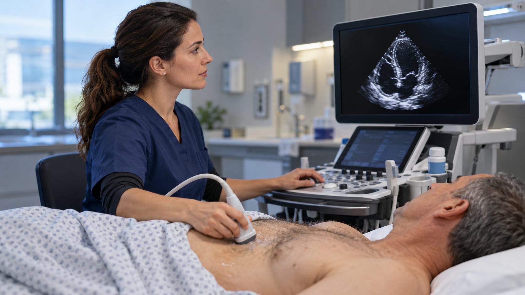
Echocardiography is not a single prediction task. It is a chain of decisions: acquiring diagnostic views, checking image quality, tracing structures, calculating measurements, interpreting findings and communicating a report. Artificial intelligence can support several of these steps, but each use has a different failure mode. A hospital should therefore evaluate the entire human–AI workflow, not only a model’s headline accuracy.
Start with the intended task
Define what the system is meant to do. Does it guide probe positioning, identify a cardiac view, trace a chamber, estimate ejection fraction, flag a possible abnormality or draft part of a report? The American Society of Echocardiography’s AI fact sheet supports responsible innovation while cautioning against inappropriate use. In practice, that means linking every output to an intended user, clinical setting and review step.
Separate image quality from measurement quality
An automated measurement can look precise even when the underlying view is incomplete or foreshortened. Acquisition quality, patient characteristics, device settings and local protocols can all affect the input. Teams should test whether the system recognizes unusable studies, exposes its confidence or limitations and lets a sonographer or physician repeat the view before a number enters the report.
Validate on the patients and devices you serve
A 2025 review in Nature Reviews Cardiology describes AI applications across acquisition, analysis and interpretation, while emphasizing the work still required for clinical integration. Local evaluation should include representative scanners, care settings and patient groups. Report performance for the actual task and examine systematic disagreement, not just average agreement.
Measure the human–AI team
A blinded randomized trial published in Nature found that an AI-guided initial assessment of cardiac function was non-inferior to a sonographer’s initial assessment in its study setting. The important implementation lesson is broader than a single result: evaluate what happens after the AI output. Track clinician edits, turnaround time, repeat acquisitions, downstream discrepancies and whether users become less attentive when an automated value appears plausible.
Keep reporting traceable
The final report should distinguish acquired observations, automated calculations and clinician conclusions. Record the model and version when appropriate, retain the source images and preserve the specialist’s ability to correct or reject a result. If an algorithm, scanner or protocol changes, assess whether the local validation still applies.
Monitor after deployment
Deployment is the start of clinical assurance, not the end. Review failed analyses, overrides, subgroup performance and drift over time. The STARD-AI reporting guideline highlights transparency, bias, generalizability, data provenance, clinical pathway integration and robustness as important elements in diagnostic AI evaluation. Those same elements provide a useful structure for post-deployment review.
This article is an editorial implementation framework, not medical advice or a claim that any specific system is suitable for diagnosis. Healthcare organizations should follow applicable regulatory requirements and professional guidance for their intended use.
- American Society of Echocardiography: AI in Echocardiography fact sheet (2025)
- Nature Reviews Cardiology: Artificial intelligence-enhanced echocardiography in cardiovascular disease management (2025)
- Nature: Blinded, randomized trial of sonographer versus AI cardiac function assessment (2023)
- Nature Medicine: STARD-AI reporting guideline (2025)