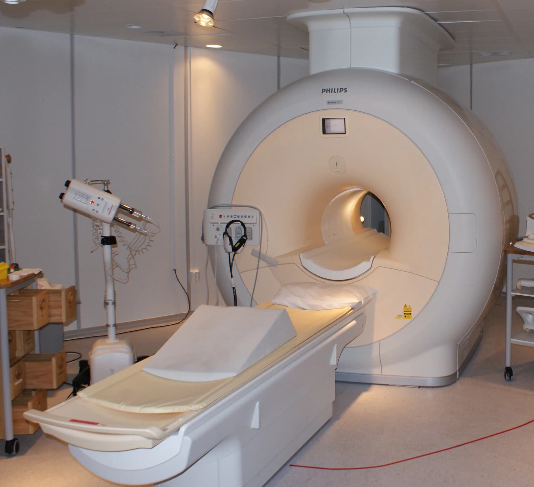
Jan Ainali / Wikimedia Commons · CC BY 3.0 · Image source
Inside UK Biobank's landmark imaging project: hitting 100,000 scanned
UK Biobank · · Source
UK Biobank’s short English-language video introducing its imaging project. It describes the research resource, not clinical authorisation for the algorithms discussed here.
Opening this video connects to YouTube and may share device information with the provider. The privacy-enhanced player is used; cookies may be used during playback. Playback does not start automatically.
A biobank holds more than sample tubes
UK Biobank announced on 15 July 2025 that it had completed imaging of 100,000 volunteers. Brain, heart, abdominal and other images can be studied alongside genetic, lifestyle and health information. Its next phase includes repeat imaging of a subset of participants to investigate change over time. That number describes the size of a research resource, not the number of people correctly diagnosed by an AI model.
The AI task: from organ boundaries to measurements
Segmentation identifies an organ’s boundaries in an image. In 2022, Kart and colleagues evaluated an automated pipeline for the liver, spleen, pancreas and kidneys using MRI data from 20,000 UK Biobank and German National Cohort participants. After appropriate post-processing, approximately nine in ten datasets had no relevant segmentation errors. Incorrect boundaries altered some organ measurements, and the authors emphasised visual quality control. This percentage is not a diagnostic accuracy rate.
Average anatomy is not one universal reference
Starck and colleagues’ 2024 study used 3,000 MRI scans to construct body atlases for six groups defined by sex and body mass index. These atlases are research references for comparing images in a shared space. The authors note that the data predominantly represent white British participants and that their atlases inherit this imbalance. Who contributed to a reference should remain visible when assessing where it can be used.
Research outputs, clinical products and authorisation
The papers discussed here evaluate research methods. Our editorial approach is to treat an organ-volume table, a physician’s report and a product’s authorised scope as separate records. Publication alone does not settle a hospital’s decision to adopt a clinical tool. This article does not determine the current regulatory status of the tools mentioned or claim authorisation for any particular product.
Editorial recommendation: preserve the trail behind each measurement
For an imaging-biobank project, we recommend recording the model version, image series, measurement unit and quality-control result alongside each measurement. Excluded examples and those needing correction should remain visible in the evaluation. These records can help teams question whether a later difference reflects biology, acquisition or processing. This educational article provides no individual diagnosis, treatment advice or evidence of clinical effectiveness for RAI products.
Sources
- UK Biobank — The world’s largest imaging project reaches milestone of 100,000 scans (15 July 2025 milestone) ↗
Accessed: 2026-10-15 - Kart et al. — Automated imaging-based abdominal organ segmentation and quality control in 20,000 participants of the UK Biobank and German National Cohort Studies (Scientific Reports, 4 November 2022) ↗
Accessed: 2026-10-15 - Starck et al. — Using UK Biobank data to establish population-specific atlases from whole body MRI (Communications Medicine, 19 November 2024) ↗
Accessed: 2026-10-15
This article is for education and information. Referencing an organization does not imply its endorsement of RAI PACS Pro. This is not a claim of clinical effectiveness for a product or model.