Image and context together
We plan to bring the hand–wrist radiograph, acquisition date and information needed for assessment into one workspace.
In development to bring bone age assessment from hand and wrist radiographs together with clinical context and clinician review.
RAI BoneAge is our bone age assessment product designed for radiology and paediatric endocrinology teams. We aim to bring the image, assessment method and report draft into one workflow that supports clinical review.
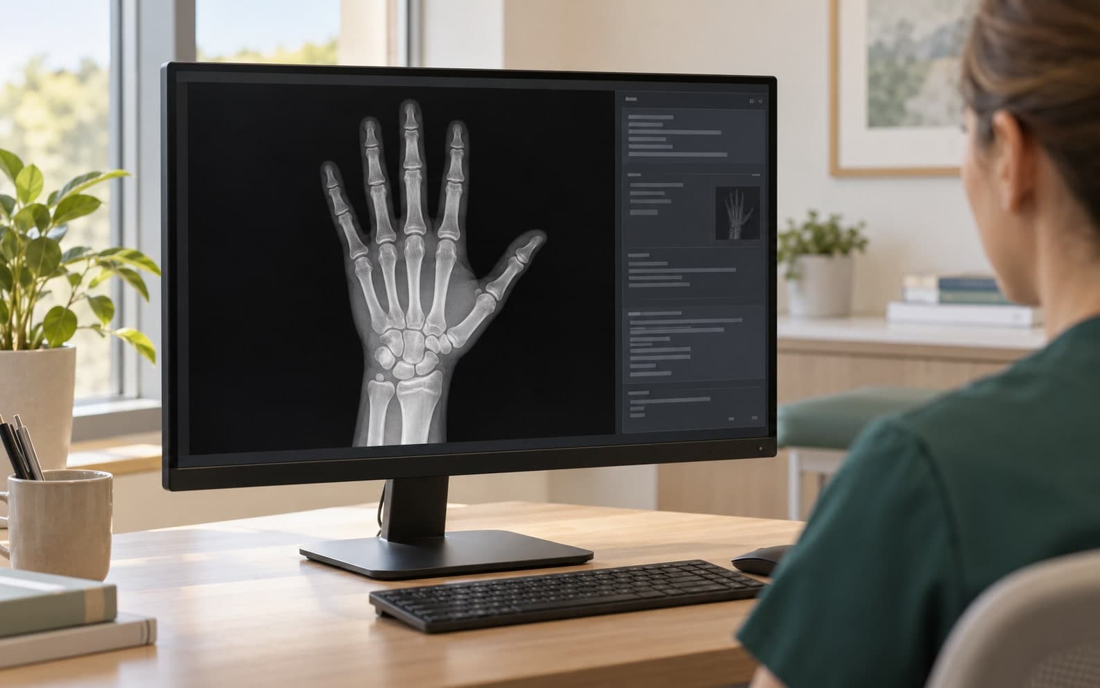
Bone age describes skeletal maturity. It is considered alongside chronological age; growth history and other clinical findings inform its interpretation. Our design gives attention to how a result is reviewed, as well as how it is presented. [1]
We plan to bring the hand–wrist radiograph, acquisition date and information needed for assessment into one workspace.
We aim to show bone age in years and months alongside chronological age, the assessment method and its limitations.
Quality and scope checks are planned at the start of the workflow, with an explained inability to assess unsuitable or unsupported images.
An editable report draft, clinician review and traceable changes. The final clinical decision remains with the clinician.
DICOM upload in Pro and study selection through PACS/VNA in Enterprise are planned. Supported images and ages will be clearly stated.
View the result, method and limitations alongside the image. Evaluate their fit with the clinical context.
Review and edit the draft. Institutional image and report transfer will follow integration validation.
These peer-reviewed studies describe the scientific background. They evaluate other developers’ systems, not the clinical validation or performance of RAI BoneAge.
In 292 children, VUNO and BoneXpert agreed closely with manual assessment; neither was significantly superior. Selected cohort; known disorders excluded.
A comparison of two artificial intelligence-based methods for assessing bone age in Turkish children: BoneXpert and VUNO Med-Bone AgeDiagnostic and Interventional RadiologyRead the studyDOI: 10.4274/dir.2024.242790In 200 radiographs, software assistance shortened two reviewers’ reading times. A retrospective study of that specific system.
Computerized Bone Age Estimation Using Deep Learning Based Program: Evaluation of the Accuracy and EfficiencyAmerican Journal of RoentgenologyRead the studyDOI: 10.2214/AJR.17.18224The BoneXpert study examined accuracy and automated self-validation. Rejecting unsuitable images is part of the assessment design.
Accuracy and self-validation of automated bone age determinationScientific ReportsRead the studyDOI: 10.1038/s41598-022-10292-ySources checked:
Our plan for RAI BoneAge makes the analysis engine version, assessment method and supported scope visible. Clinical validation, local conditions of use and institutional integration will be assessed separately.
The product information page is available; the calculation service and clinical use are not yet open. Supported scope and access details will be shared when ready.
Greulich–Pyle and Tanner–Whitehouse methods inform our product evaluation. The offered method will depend on the selected analysis engine and validation results, and will be stated in each report.
The initial scope is bone age assessment and a clinician-reviewed report. Adult height prediction and a bone health index are being considered for later phases with separate validation.
No. The publications examine the field and other systems. RAI BoneAge needs its own clinical performance evaluation; no RAI accuracy rate or regulatory clearance is claimed here.