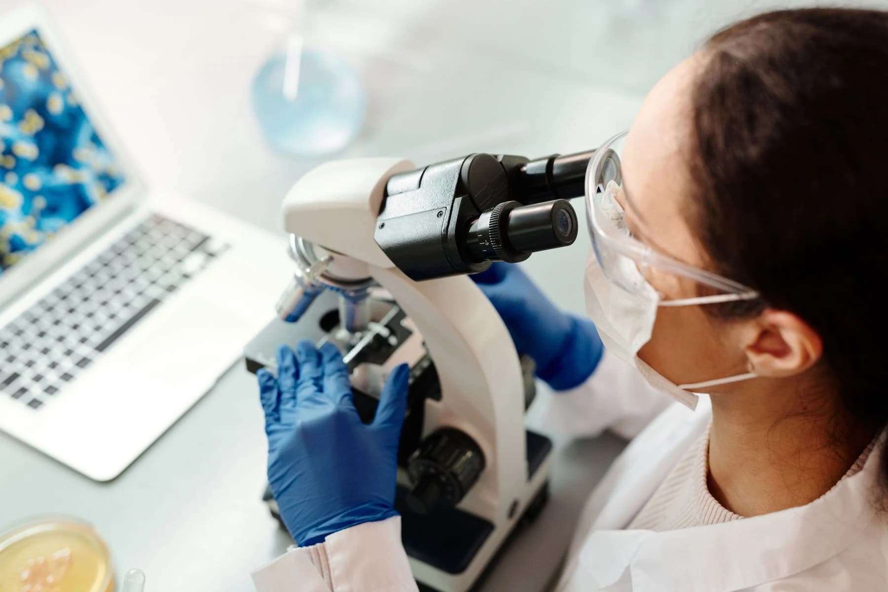
Edward Jenner / Pexels · Pexels License · Image source
Digital Pathology Imaging in Cancer Clinical Trials and Research — Day 1
National Cancer Institute (NCI) · · Source
Full workshop recording in English, approximately 6 hours 37 minutes. Event: 6 March 2024. The date above is the upload date shown by NCI’s player (29 April 2026).
Opening this video loads NCI’s Vbrick player. Your IP address and device information may be shared with the provider, which may use cookies. Playback does not start automatically.
Research: learning without marking every cell
Campanella and colleagues’ 2019 study evaluated deep learning using a dataset of 44,732 whole-slide images from 15,187 patients. Training used reported diagnoses rather than pixel-level annotations, examining prostate cancer, basal cell carcinoma and breast cancer metastases in lymph nodes. These are research findings in specified datasets, not proof of equivalent performance in every laboratory or benefit in routine patient care. The publication discloses financial or intellectual-property relationships between some researchers, their institution and Paige.AI.
Regulatory record: a defined task and device configuration
FDA’s DEN200080 decision summary describes a specific adjunctive use for Paige Prostate: helping pathologists re-examine suspicious areas in H&E-stained prostate needle biopsies after their initial review. It specifies the Philips Ultra Fast Scanner and Paige FullFocus viewer; the AI output must not serve as the primary diagnosis. This US record is not blanket authorization for pathology AI, other scanners or other tissue types, nor does it establish permission to use a product in Europe or Turkey.
Implementation: a workflow beyond the scanner
NCI’s digital pathology workshop addresses image quality, standard operating procedures, interoperability, de-identification and access governance together. Purchasing a scanner alone does not establish the necessary infrastructure. This source is a workshop summary and implementation discussion, not an assessment of a specific AI product’s clinical effectiveness.
Editorial interpretation: questions for local evaluation
A pilot can define the model’s task and version, specimen preparation, scanner compatibility and responsible reviewer together. Recording when a suggestion is rejected and which images cannot be assessed may be more informative than tracking a success rate alone. A research paper, a regulatory record and local validation are not interchangeable. This article is neither a new authorization announcement nor personal diagnostic or treatment advice.
Sources
- Campanella et al. — Clinical-grade computational pathology using weakly supervised deep learning on whole slide images (Nature Medicine, 2019) ↗
Accessed: 2026-10-05 - FDA — Paige Prostate, De Novo Decision Summary, DEN200080 ↗
Accessed: 2026-10-05 - NCI — Workshop on Digital Pathology Imaging (DPI) in Cancer Clinical Trials and Research (2024; page updated 2026) ↗
Accessed: 2026-10-05
This article is for education and information. Referencing an organization does not imply its endorsement of RAI PACS Pro. This is not a claim of clinical effectiveness for a product or model.