What task does AI perform in an image?
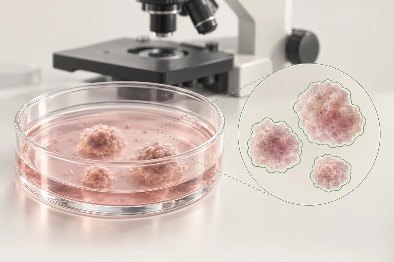
Organoids are three-dimensional cellular structures grown in a laboratory. The 2023 OrgaExtractor study used deep learning to outline colon organoids. This task, segmentation, divides images into meaningful regions.
Why include conventional methods in the comparison?
The 2026 OrganoSeg2 study compared a method requiring no deep learning with AI tools. Evaluation across six organoid types suggests testing method selection against the imaging conditions.
Editorial assessment: define the measurement question first
Our recommendation for a research team is to specify the output before choosing a model: counting individual structures, measuring their area or tracking the same structure over time? Then connect acceptance criteria to that task. A small boundary error and counting two neighboring organoids as one are different problems. An overall performance score should not conceal that distinction.
How could an independent laboratory evaluation be planned?
Our editorial recommendation is to reserve culture batches not used in training for evaluation, record the microscope and acquisition settings, and examine small or touching structures separately. Report expert corrections and unusable images as well. Explaining how failed examples are handled makes an evaluation more informative than showing only a few successful examples.
How do measurement, biological response and patient outcome differ?
Our proposed evaluation keeps three questions separate: was the structure outlined correctly; is the measurement associated with the biological endpoint selected for the study; does that information improve decisions or outcomes for a patient? Answering the first does not answer the others. Before calling an area change “successful treatment,” for example, identify the independent evidence used to test that interpretation.
What may still be missing before clinical use?
The tools discussed here are research examples. They should not be treated as evidence of clinical authorization for a product or effectiveness of RAI solutions. Institutional assessment should consider intended use, human review, data governance and product-specific validation documents separately. This article is not intended to guide personal treatment selection.
Sources
This article is for education and information. Referencing an organization does not imply its endorsement of RAI Health Pro. This is not a claim of clinical effectiveness for a product or model.